Safety & Convenience
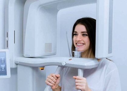
As in a standard CT scan, a cone beam scanner uses x-rays but at 10% to 20% of the dose. Time within the scanner whilst operational is quite brief.
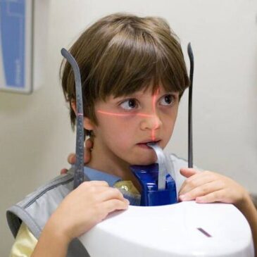
Whilst consideration should always be given to exposure, particularly for children, a reduced dose is a notable safety advantage.
As the scanner rotates around you, the images gathered are quickly processed into 3D visualisations. Positioning you takes just minutes, the scans themselves seconds and complete images are produced in a few more minutes.
Scanning is entirely painless, no medical aftercare is needed, you will be able to eat, drink and carry on with your day as you wish. At the same time, your consultant has immediately available information on your condition.
Medical Advantages
Soft tissue and bone can be looked at simultaneously, giving the detail needed for diagnosis. The images help form a basis for your consultation, a first step in dealing with medical issues and restoring your health
Information provided is equally valuable for treatment, surgical, or restorative planning. The scanner is available to all departments and if using this makes sense for your needs, a complete explanation of procedure will be given.
The system’s targeting and automated information processing then get to work. Creating 3D data which can be manipulated to improve visualisation, or imported into other software for detailed study. The comprehension and level of precision available are recognised as field leading.
Research On Cone Beam Tomography
A cone beam computed tomography (CBCT) scan can be isolated and manipulated, more or less in real time. This has led researchers and professional bodies to state that CBCT is now the gold standard for maxillofacial care.
Others would say that MRI scans are still better for certain conditions and this can be true, although there is misunderstanding.
MRI scans tend to be seen as the best route for temporomandibular joint osteoarthritis. In a May 2023 paper, a team set out to compare MRI effectiveness to CBCT, concluding that CBCT improved results by around 40%.
There are studies which go the other way, such as one which looked at soft tissue analysis in the mandibular nerve. There is however a caveat, the study in question was carried out in 2012 and the position has changed.
Significant software advances have improved signal to noise ratio on CBCT scans and increased visual contrast. Cone beam computed tomography systems were originally designed to provide hard tissue visualization but are now far more versatile.
Into The Future
Governments have set up research programs to study the effectiveness of different systems, with larger alternatives to CBCT still in demand but expensive.
The chances are they will opt for increased use of smaller scale devices, especially for maxillofacial units. They are ideal for studying teeth, jaws, TMJ, airways, sinuses and surrounding anatomical structures.
Interactive 3D images speed up diagnosis, planning and treatment, to the benefit of patients. They are convenient, make a difference to how long discomfort lasts and to outcome.
The benefit to patients is evident, improved medical understanding which they can physically see. No machine can replicate the skills of an experienced consultant but knowing the problem is understood brings peace of mind.
Artificial intelligence has the potential to take CBCT scans further, through algorithmic identification of anomalies. Finer fields of view are being developed, along with other image enhancements.
Our team are well trained on CBCT scanning and watch developments closely. You can be assured we will remain at the forefront of cone beam scanning and the advantages this brings for all forms of maxillofacial treatment.


